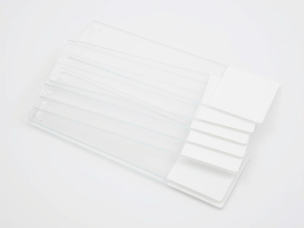
Treated with special reagents and processes, they deliver durable and stable adhesive performance.
Product Description
Long-lasting Strong Adhesion
Treated with specialized reagents and processes, it delivers durable, stable adhesion that firmly secures tissue sections and significantly reduces the risk of sample detachment in subsequent steps.
Premium Glass Substrate
Made of high-transparency, structurally dense and stable glass with excellent chemical resistance. The adhesion layer bonds tightly to the substrate, ensuring overall reliability throughout experimental procedures.
Minimal Background Interference
The substrate exhibits no autofluorescence, minimizing non-specific background signals to provide a clear imaging baseline for high-precision assays such as FISH and ISH.
Broad Experimental Applicability
Recommended for use across a wide range of preparation techniques including immunohistochemistry (IHC), frozen sections, liquid-based cytology (LBC), and routine H&E staining, with high compatibility.
Suitable for Challenging Samples
Particularly suited to sections of difficult-to-adhere tissues such as fatty or calcified specimens, effectively improving preparation success rates and stability for these sample types.
ViePath™ Adhesive Slides are treated with specialized reagents and processes to deliver durable, stable adhesion performance, ensuring tissue sections remain firmly attached and effectively reducing the risk of sample detachment during subsequent processing steps.
The product uses a high-transparency glass substrate with a dense, stable structure and excellent chemical resistance. The adhesion layer bonds tightly to the substrate, which exhibits no autofluorescence, minimizing background signal interference and making it particularly suitable for FISH and ISH applications.
Recommended for use in immunohistochemistry (IHC), frozen sections, liquid-based cytology (LBC), and routine H&E staining preparation, including difficult-to-adhere samples such as fatty or calcified tissues.
Precautions for Use: · Conduct a preliminary validation test before first use. · Improper tissue pre-processing or inherently poor tissue adhesion may increase the risk of slide detachment. · Avoid contact with the functional adhesion area during operation to prevent contamination that may compromise adhesion performance. · Strongly alkaline environments may cause the colored coating to peel off. · Store in dry, room-temperature conditions. Use promptly after opening.
Product No.
Product Specification
Packing
MS181205
75×25mm, 1.0–1.2mm thickness, 45° corners, polished edges. Positively charged surface with white painted frosted end.